
You want clear answers about your teeth. Cone beam imaging gives your dentist a three dimensional view of your mouth, jaw, and roots. This clear picture helps find problems that regular X rays can miss. It also helps plan safe treatment. You face less guesswork and fewer surprises. This matters when you need implants, root canals, or Wantagh dental crowns. It also matters when you feel pain, but standard tests show nothing. With cone beam imaging, your dentist can see bone loss, hidden fractures, and nerve paths. That means better choices, fewer repeat visits, and more peace of mind. You gain a plan that fits your mouth, not a rough estimate. You deserve care based on sharp facts, not blurry images. Cone beam imaging provides that sharp view and supports stronger, safer decisions about your care.
What Cone Beam Imaging Is
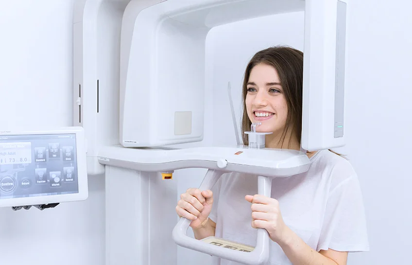
Cone beam imaging is a type of three dimensional X ray. The machine circles your head and collects many small images. Then a computer joins these images into one detailed model of your teeth, jaw, and joints.
You stand or sit still. The scan takes less than one minute. You do not feel pain. You keep your mouth closed or bite on a small piece of plastic. The machine does the work.
Traditional dental X rays show flat pictures. Cone beam imaging shows height, width, and depth. You see the full shape of roots, bone, and spaces. Your dentist can turn the image and look from many angles.
Why This Matters For Your Health
A clear picture can protect your health. A missed problem can grow and cause stronger pain or tooth loss. Early detection stops that chain.
With cone beam imaging, your dentist can:
- Find infections at the tip of roots before they spread
- Measure bone thickness before placing an implant
- Check sinus spaces and jaw joints when you have pressure or popping
The U.S. Food and Drug Administration explains cone beam imaging and its use in dental care. You can read about how it collects data and how dentists use it to plan treatment. This helps you ask clear questions and feel calm before a scan.
How Cone Beam Imaging Compares To Regular X Rays
Both tools use X-rays. They serve different purposes. Regular X-rays work well for routine checks. Cone beam imaging works best when your case is complex or when past images do not explain your symptoms.
| Feature | Regular Dental X Rays | Cone Beam Imaging |
|---|---|---|
| Type of image | Flat two dimensional picture | Three dimensional model |
| View of roots and nerves | Limited detail | Clear path and shape |
| Use for implants | Rough estimate of bone | Exact height, width, and angle |
| Use for hidden fractures | Can miss small cracks | Better view of fine lines |
| View of jaw joints | Often not covered | Shows joints and nearby bone |
| Typical use | Routine exams and cavities | Complex planning and tough cases |
Both tools can fit into safe care. Your dentist chooses the right one for your needs. You can ask why a cone beam scan is suggested and how it will change your plan.
Better Planning For Implants And Crowns
Implants and crowns stay in your mouth for many years. Poor planning can lead to pain, lost work, or broken teeth. Cone beam imaging lowers these risks.
For implants, cone beam imaging helps your dentist to:
- Measure bone height and width where the implant will sit
- See how close nerves and sinus spaces are to the site
- Choose the right size and angle for the implant
For crowns, the scan can:
- Show cracks under old fillings
- Reveal hidden decay near the root
- Guide treatment if a root canal is also needed
This level of planning can protect healthy teeth near the treatment site. It also reduces the chance that work needs to be redone.
Support For Root Canals And Jaw Pain
Root canals work best when the dentist can see each root and canal. Some teeth have more branches than expected. A flat X-ray can hide a thin canal. A missed canal can keep infection alive.
Cone beam imaging can help your dentist to:
- Count all roots and canals before starting
- Locate curved or narrow canals
- Check healing after treatment
Jaw pain and popping can feel confusing. You may feel pain near your ear or in your head and not know the cause. Cone beam imaging shows the jaw joints, nearby bone, and bite. Your dentist can see if the joint shape or tooth contact adds strain.
Radiation Dose And Safety
Many people fear X-rays. That fear is natural. You want to protect your body and your children. Information helps guide calm choices.
The National Institutes of Health shares data on dental cone beam imaging and dose levels. These reports show that cone beam scans use more radiation than a single small dental X-ray, yet often less than a full medical CT of the head.
Your dentist can lower your exposure by:
- Ordering cone beam scans only when they change treatment
- Using the smallest scan size that still shows what is needed
- Placing a lead apron and thyroid collar when appropriate
You can ask how the dose compares to other common imaging tests. You can also ask what decisions the scan will support. When a scan prevents failed treatment or surgery, the benefit can outweigh the risk.
Questions To Ask Your Dentist
You have the right to understand each step of your care. Before a cone beam scan, you can ask:
- Why do I need this scan today
- What can this show that regular X-rays cannot show
- How will the results change my treatment plan
- What is the radiation dose compared with my last set of X-rays
- Can you show me the images and explain what you see
Clear answers build trust. They also help you feel calm during care. When you see your own three-dimensional images, you can share in each choice about your mouth.
Using Cone Beam Imaging Wisely
Cone beam imaging is a strong tool. It is not needed for every visit. You gain the most when it is used to answer a hard question or to plan complex care such as implants, root canals, or crowns.
With careful use, cone beam imaging can:
- Find problems early
- Steer safer treatment steps
- Reduce repeat work and visits
You deserve care that respects your time, money, and health. A clear three-dimensional view helps your dentist respect all three. When you understand why cone beam imaging is used, you can move through treatment with more trust and less fear.